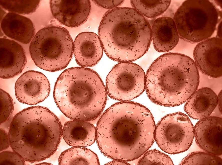
Protein homeostasis stands as one of the most critical yet overlooked processes in our cells. This delicate balancing act uses much of the cell’s energy. Cells must dedicate much of their ATP resources to keep proteins functioning properly. Any disruption in this balance can have devastating effects.
This piece explores the meaning of homeostasis in cells, with special focus on protein homeostasis in health and disease. We’ll learn how this complex system maintains cellular protein homeostasis through synthesis, folding, trafficking and degradation.
How protein homeostasis supports cellular health
Protein homeostasis at the cellular level works like a complex balancing act between creation and destruction. This dynamic process includes protein synthesis, folding, trafficking and degradation. These activities use up 66% of cellular energy resources as ATP equivalents.
Why balance between synthesis and degradation is critical
Protein synthesis and degradation are the life-blood of cellular functionality. Cells immediately break down about one-third of newly made proteins. This process might seem wasteful but serves a crucial purpose. It helps cells adjust their proteome faster to changing conditions and remove harmful misfolded proteins.
Cells can suffer damage from even slight imbalances between synthesis and degradation rates. Non-dividing cells need precise coordination to keep protein levels steady despite continuous turnover. Cycling cells must double their protein content during each cell cycle. Chemical feedback alone can’t achieve this balance. The process needs sophisticated regulatory mechanisms between protein manufacture on ribosomes and breakdown in proteasomes.
Homeostasis and cells: a dynamic equilibrium
A network of approximately 1400 proteins maintains proteostasis by coordinating synthesis, folding and degradation activities. This network exists in all subcellular compartments and uses specialized systems based on protein types and locations.
Cells activate specific pathways like endoplasmic reticulum-associated protein degradation (ERAD) and unfolded protein response (UPR) when proteins don’t fold correctly. The process of autophagy targets aggregated proteins and breaks them down through lysosomes. These quality control mechanisms work non-stop to keep the proteome functional despite environmental stressors.
How proteostasis affects organ function and longevity
The protein homeostasis network’s efficiency impacts both healthspan and lifespan by a lot. Our bodies become less able to maintain protein homeostasis as we age. This decline leads to tissue deterioration and connects to many age-related disorders, from neurodegeneration to cancer.
Research shows that better proteostasis capacity can extend lifespan in species of all types. Scientists found that higher levels of molecular chaperones like HSP70 and HSP16 help increase longevity. Dietary changes like caloric restriction and intermittent fasting support proteostasis. These changes activate pathways that control protein quality. This suggests that maintaining protein homeostasis could be key to healthy aging.
What causes protein homeostasis to break down
Cells need constant alertness to maintain their protein balance against many disruptive forces. Life presents multiple challenges to this delicate equilibrium that push cells toward proteostasis collapse.
Environmental and oxidative stress
Cellular metabolism creates reactive oxygen species (ROS) as an unavoidable byproduct. These reactive molecules damage proteins through oxidation in two ways: reversible modifications that cells can repair and irreversible damage that permanently changes protein structure. The original oxidation might only affect function slightly, but additional oxidative damage guides proteins to unfold. This exposes hydrophobic regions and creates insoluble aggregates that resist degradation. Temperature changes, pressure shifts and toxic exposure put extra stress on proteostasis by overwhelming the cell’s defense systems.
Genetic mutations and misfolding
Mutations occurring in protein-coding sequences usually make proteins more likely to misfold. These changes can destabilize the protein’s structure and cause improper folding, early degradation or toxic clumping. Mutations affect more than just protein stability, they also disrupt translation rhythm. Codon usage patterns create strategic pauses during protein synthesis and mutations that alter these patterns can impair folding by a lot without changing translation speed.
Aging and reduced chaperone activity
The body’s chaperone availability and function decline steadily with age. Research shows that levels of key chaperones like Hsc70 and Hsp90 drop by a lot in aged tissues. The proteasome becomes less active due to structural changes, fewer proteasome subunits and oxidative damage to the protein-breaking machinery. These age-related problems create a dangerous cycle. The reduced number of working chaperones results in more misfolded proteins that overwhelm the already weakened protein quality control systems.
Impact of chronic inflammation
Chronic inflammation, common in aging, creates a hostile environment for protein homeostasis. The inflammatory process generates more reactive species that damage proteins and strain cellular repair systems. This creates a harmful cycle: disrupted proteostasis triggers inflammation, which makes protein damage worse. Brain inflammation especially reduces cells’ ability to handle proteotoxic stress, making the brain vulnerable to proteostasis breakdown.
How to support protein homeostasis: lifestyle and supplements
Research shows that lifestyle changes and specific supplements can improve cellular protein quality control systems by a lot. This offers practical ways to maintain optimal proteostasis.
Caloric restriction and fasting
Caloric restriction (CR), reducing daily calorie intake by 30-50% without causing malnutrition, helps us live healthier and keeps stress response working well as organisms age, according to studies. Our body clears damaged proteins better because this method activates autophagy by blocking mTOR signaling. Brief periods of fasting work just as well, even a single early-life fast can improve HSF-1 activity and help maintain proteostasis capacity. Research shows that fasting for just 12 hours gets more proteasome activity going in mouse liver cells. This proves that short periods without food might be enough to kick-start protein quality control mechanisms.
Exercise and its effect on proteostasis
Exercise provides a powerful way to maintain proteostasis. Our muscle cells’ protein turnover changes a lot during exercise and these changes last up to 72 hours afterward. Human muscle cells show much higher molecular signs of protein breakdown after intense biking. Our skeletal muscles adapt to different types of training because exercise boosts muscle protein synthesis rates. Physical activity makes this happen by triggering cellular stress responses that increase chaperones and proteolytic pathways needed for protein quality control.
Supplements that support chaperone function
Heat shock proteins (HSPs) protect cells from proteotoxic stress. HSP inducers (HSPis) are natural or synthetic compounds that boost HSP expression by activating heat shock transcription factors. These include:
- Geranylgeranylacetone (GGA): this safe compound boosts HSP levels through HSF1 activation and doctors have used it as an antiulcer drug since 1984;
- Pro-Tex®: a liquid form from Opuntia ficus indica that triggers HSPs;
- Amygdalin: a natural compound found in apricots and plums;
- Pyrazole derivatives: these get HSF1 working in human cells.
mTOR inhibitors and autophagy activators
The mechanistic target of rapamycin (mTOR) controls proteostasis and blocking it mimics what happens when nutrients are low, which triggers autophagy. Rapamycin specifically blocks mTOR and helps organisms from yeast to mammals live longer. Lower TOR signaling helps nematodes, flies and yeast live longer too. Scientists have found other autophagy activators besides rapamycin. Metformin helps aging heart muscle cells work better and various mTOR blockers show good results in mice with Alzheimer’s.
Natural compounds like resveratrol and spermidine
Resveratrol and spermidine are natural compounds that strongly affect proteostasis. Grapes and red wine contain resveratrol, which triggers autophagy through SIRT1 activation. Spermidine, found in citrus fruits and soybeans, boosts autophagy without using SIRT1. Both help various species live longer. Spermidine works by changing eIF5A and blocking histone acetyltransferase P300. These compounds work even better together, they can trigger autophagy at doses that wouldn’t work alone.
Emerging therapies targeting proteostasis
Scientists keep finding new ways to target proteostasis. They’ve found a nucleolar complex that plays a key role in keeping cells healthy. Blocking this complex reduces toxic effects from Alzheimer’s-causing proteins. Animal studies show promise with gene therapy that increases autophagy-related proteins like Beclin-1. Ketogenic approaches look promising too, as ketone bodies control autophagic flux and chaperone-mediated autophagy. R-βHB directly stops amyloid-β from being toxic in rat brain cells, while both ketogenic diets and added ketones reduce brain plaque in several mouse models, according to studies.
Protein homeostasis is the life-blood of cellular health and has major implications for longevity and disease prevention. Caloric restriction and intermittent fasting trigger autophagy pathways, while regular exercise boosts protein turnover and quality control mechanisms. It also helps that natural compounds like resveratrol and spermidine provide promising benefits by inducing autophagy through different molecular pathways.
The genetic control of individual proteostasis networks points to highly tailored approaches for maintaining cellular protein balance.


