Scientists have discovered that lipofuscin and aging have a deeper connection than they originally thought. This autofluorescent lipopigment builds up in post-mitotic cells, especially in the brain’s, heart’s and skin’s cells. It acts as a universal aging marker in organisms of all types, including humans.
Our understanding of lipofuscin gives us valuable clues about extending healthy lifespans in the complex field of cellular aging.
What is lipofuscin?
The yellow-brown substance lipofuscin stands out as one of the most common markers of cellular aging. Scientists call it “dark lipid” (its Latin name) and this fascinating cellular material builds up throughout life when cells can’t process their waste completely.
Definition and composition of lipofuscin pigment
Light microscopy reveals lipofuscin as yellow-brown granules approximately 0.5 to 3 μm in diameter. This complex substance contains oxidized proteins and lipids such as triglycerides, free fatty acids, cholesterol and phospholipids. A small amount comes from carbohydrates and metals, mostly iron.
Lipofuscin’s most unique feature is its autofluorescent property. The substance glows with a golden-yellow to orange-red fluorescence under ultraviolet or blue light, spanning a broad spectrum (400-700nm). Each tissue type shows a different fluorescent pattern because of lipofuscin’s varied nature.
Cells cannot break down lipofuscin once it forms. The substance creates covalently cross-linked combinations that resist normal cellular cleanup processes like the ubiquitin-proteasome system. Its insoluble properties also prevent cells from removing it through exocytosis.
Where it accumulates in the body
Lipofuscin builds up mostly in postmitotic cells, cells that rarely or never divide, especially those with high metabolic activity. The most important accumulations happen in:
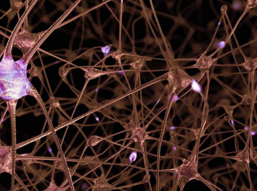
- Neurons throughout the brain and spinal cord;
- Cardiac muscle cells (myocytes);
- Retinal pigment epithelium cells;
- Liver cells (hepatocytes);
- Adrenal cortical cells.
The human brain shows specific patterns of lipofuscin distribution, especially in larger neurons. The core team found substantial age-related buildup in motor neurons of the spinal cord’s anterior horn, cells in the inferior olivary nucleus and neurons in the cerebral cortex. Some parts of the hypothalamus seem resistant to lipofuscin accumulation.
Why it’s called the ‘aging pigment’
Several compelling features earned lipofuscin its nickname “aging pigment.” The substance builds up almost linearly with age in organisms of all sizes. Human brain cells show small amounts of lipofuscin granules in early childhood and these levels steadily increase between the second and ninth decades of life.
Scientists associate slower lipofuscin buildup with longer lifespans. Research comparing long-lived and short-lived monkey species revealed similar lipofuscin amounts in heart muscle cells at the same relative age (compared to maximum lifespan).
Motor neurons in humans over one hundred years old can contain up to 75% lipofuscin in their total cell volume, according to research. This extensive accumulation makes lipofuscin one of the most reliable and measurable indicators of cellular aging.
How lipofuscin builds up over time
The biological events that create lipofuscin in cells follow a complex pattern. This “aging pigment” builds up through oxidation processes. The way it accumulates tells us a lot about how cells age.
Role of oxidative stress and mitochondrial damage
Oxidative stress kicks off lipofuscin formation. Research shows that pro-oxidants make lipofuscin accumulate faster, while antioxidants slow down this process. Inside cells, oxidized proteins and lipids link together through aldehyde bridges. These new structures resist normal breakdown processes.
The mitochondrial-lysosomal axis theory of aging suggests damaged mitochondria create most lipofuscin. These cellular powerplants make reactive oxygen species (ROS) during normal metabolism. Damaged mitochondria produce too much superoxide and hydrogen peroxide, which leads to more lipofuscin. Studies show that animals living longer have mitochondria that make nowhere near as much hydrogen peroxide as short-lived species.
Decline in lysosomal and autophagy efficiency
Our cells’ recycling systems become less efficient with age. Lysosomes, once known as “roving garbage disposal units,” get stuffed with lipofuscin that won’t break down. This buildup creates a harmful cycle where lysosomes loaded with lipofuscin can’t help with autophagy anymore.
Research shows that autophagy, the process that removes damaged cell parts, drops with age. Lipofuscin makes this worse by blocking lysosomal enzymes.
Why long-lived cells are most affected
Neurons, heart muscle cells and retinal pigment epithelium cells carry the heaviest lipofuscin load. These cells can’t divide to spread out lipofuscin like other cells do, so it keeps building up.
The motor neurons of people over one hundred years old can contain up to 75% lipofuscin in their cell fluid. This huge buildup happens because these cells stay in the G0-phase and can’t divide to share the material between new cells.
Lipofuscin and its role in aging and disease
Lipofuscin does more than just mark aging, it plays an active part in how age-related diseases develop and spread through many organ systems. This substance might actually cause pathological changes rather than simply indicating them.
Lipofuscin in Alzheimer’s and Parkinson’s disease
Neurons in the prefrontal cortex and hippocampus of Alzheimer’s patients build up lysosomes filled with lipofuscin aggregates. These lipofuscin deposits end up in extracellular spaces after neurons die, and scientists often find them near senile β-amyloid plaques. Studies comparing Alzheimer’s patients with people of similar age showed no major differences in lipofuscin levels in some brain areas, which suggests a more complex relationship.
Scientists have found stronger links to Parkinson’s disease. Lipofuscin granular aggregates seem to directly cause the death of dopaminergic neurons. Research shows that only neurons containing lipofuscin or neuromelanin become more likely to develop pathological changes, including α-synuclein Lewy body deposits. Scientists have found α-synuclein as part of lipofuscin deposits in the substantia nigra of Parkinson’s patients.
Impact on retinal cells and macular degeneration
Age-related macular degeneration (AMD) ranks as the top cause of vision loss in older people, and it clearly links to lipofuscin buildup. Lipofuscin fills the retinal pigment epithelium, absorbs blue light, and creates toxic effects through reactive oxygen intermediates. A2E, a major lipofuscin fluorophore, helps create light-induced oxidative stress and breaks down membrane structure.
Is lipofuscin a cause or a marker of aging?
Scientists used to think lipofuscin was just a harmless byproduct of aging. New evidence suggests it actively contributes to cell deterioration. Pure lipofuscin kills cells at surprisingly low concentrations (LC50 = 5.0 μg/mL) through pyroptosis-like pathways. Some neurons packed with lipofuscin resist age-related degeneration, which makes the picture more complex.
How it disrupts cellular recycling and function
Lipofuscin interferes with cells in several ways. The substance blocks the ubiquitin-proteasome system by binding to proteolytic enzymes. It reduces what lysosomes can do by trapping enzymes. The redox-active surface of lipofuscin speeds up the Fenton reaction, which creates free radicals that damage cell parts. These changes lead to less efficient autophagy, more oxidative stress and eventually cell death.
Can we reduce or prevent lipofuscin accumulation?
Scientific breakthroughs have changed what we thought about lipofuscin accumulation. Scientists believed this aging pigment would stay permanently once it formed. New evidence suggests we might slow down or reduce lipofuscin buildup.
Supplements under study: spermidine, melatonin, CoQ10
Scientists are testing several compounds that could help fight lipofuscin accumulation. Spermidine, which naturally occurs in fresh peppers, wheat germ and soy products, shows remarkable anti-aging properties. Long-term spermidine supplementation (1.2 mg/day) is safe and works well for elderly people, according to studies. This polyamine works like caloric restriction through autophagy pathways and helps boost memory in older adults who notice cognitive decline.
Coenzyme Q10 (CoQ10) helps produce cellular energy and acts as a powerful antioxidant. Research shows that using CoQ10 with melatonin for 4 weeks reduced lipofuscin in rat hippocampus. Melatonin helps in two ways, it improves sleep quality and lowers cortisol levels, which might ease oxidative stress that creates lipofuscin.
Lifestyle interventions: fasting, caloric restriction
Caloric restriction stands out as one of the best ways to reduce lipofuscin. Studies show that mice fed 2g/day for 12 months learned faster, performed better, and had much less lipofuscin in their hippocampal and frontal cortex neurons. Severe caloric restriction (52% of normal diet) slows lipofuscin buildup, while moderate restriction (25%) barely makes a difference, according to research.
The key benefits of caloric restriction need longer fasting periods. A short 7-day fast substantially reduced lipofuscin in marine snails’ hepatopancreatic cells. This suggests the mechanism works across different species.
Experimental therapies and future directions
Drug-based approaches look promising. Rapamycin boosts autophagy and reduces lipofuscin buildup and aging in rat heart cells. Remofuscin (soraprazan) reverses lipofuscin accumulation in aged human retinal pigment epithelium cells. This could help treat age-related macular degeneration.
Lipofuscin does much more than just mark aging in our cells. This undegradable cellular waste plays an active role in age-related decline through several mechanisms. Lipofuscin builds up mainly in long-lived, post-mitotic cells and gradually impairs cellular function by disrupting lysosomal efficiency and autophagy processes.